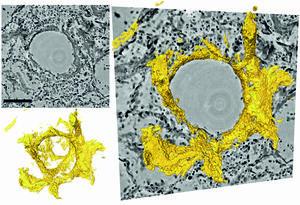
Sections through the three-dimensional reconstruction volume (upper left, grey) around a pulmonary alveolus with hyaline membrane (lower left, yellow). On the right, the images are superimposed. In the center is the air bubble (alveolus). The electron density is represented by different shades of grey. On the inside of the air bubble is a layer of proteins and dead cell residues, the "hyaline membrane". This deposit, which can be represented in its three-dimensional structure for the first time by the new method, reduces the gas exchange and leads to respiratory distress. Courtesy of T Salditt, M Eckermann
Physicists at the University of Göttingen, together with pathologists and lung specialists at the Medical University of Hannover, have developed a three-dimensional imaging technique that enables high resolution and three-dimensional representation of damaged lung tissue following severe Covid-19. Using a special X-ray microscopy technique, they were able to image changes caused by the coronavirus in the structure of alveoli (the tiny air sacs in the lung) and the vasculature. The results of the study were published in the research journal eLife.
In severe Covid-19 disease, the researchers observed significant changes in the vasculature, inflammation, blood clots and "hyaline membranes," which are composed of proteins and dead cells deposited on the alveolar walls, which make gas exchange difficult or impossible. With their new imaging approach, these changes can be visualized for the first time in larger tissue volumes, without cutting and staining or damaging the tissue as in conventional histology. It is particularly well suited for tracing small blood vessels and their branches in three dimensions, localizing cells of the immune systems which are recruited to the inflammation sites, and measuring the thickness of the alveolar walls. Due to the three-dimensional reconstruction, the data could also be used to simulate gas exchange.
"Using zoom tomography, large areas of lung tissue embedded in wax can be scanned enabling detailed examination to locate particularly interesting areas around inflammation, blood vessels or bronchial tubes," says lead author professor Tim Salditt from the Institute of X-ray Physics at the University of Göttingen. Since X-rays penetrate deep into tissue, this enables scientists to understand the relation between the microscopic tissue structure and the larger functional architecture of an organ. This is important, for example, to visualize the tree of blood vessels down to the smallest capillaries.
The authors foresee that this new X-ray technique will be an extension to traditional histology and histopathology, areas of study which go back to the 19th century when optical microscopes had just become available and pathologists could thereby unravel the microscopic origins of many diseases. Even today, pathologists still follow the same basic steps to prepare and investigate tissue: chemical fixation, slicing, staining and microscopy. This traditional approach, however, is not sufficient if three-dimensional images are required or if large volumes have to be screened, digitalized or analysed with computer programs.
Three-dimensional imaging is well known from medical computerized tomography (CT). However, the resolution and contrast of this conventional technique are not sufficient to detect the tissue structure with cellular or sub-cellular resolution. Therefore, the authors used "phase contrast", which exploits the different propagation velocities of X-rays in tissue to generate an intensity pattern on the detector. Salditt and his research group at the Institute for X-ray Physics developed special illumination optics and algorithms to reconstruct sharp images from these patterns, an approach which they have now adapted for the study of lung tissue affected by severe progression of Covid-19. The Göttingen team could record lung tissue at scalable size and resolution, yielding both larger overviews and close-up reconstructions. Depending on the setting, their method can even yield structural details below the resolution of conventional light microscopy. To achieve this, the researchers used highly powerful X-ray radiation generated at the PETRAIII storage ring of the German Electron Synchrotron (DESY) in Hamburg.
As was the case when the modern microscope was invented 150 years ago, significant progress has resulted from collaboration between physicists and medical researchers. The interdisciplinary research team hopes that the new method will support the development of treatment methods, medicines to prevent or alleviate severe lung damage in Covid-19, or to promote regeneration and recovery. "It is only when we can clearly see and understand what is really going on, that we can develop targeted interventions and drugs," adds Danny Jonigk of Medical University Hannover, who led the medical part of the interdisciplinary study.
Reference: M. Eckermann, J.Frohn, M. Reichardt, M. Osterhoff, M. Sprung, F. Westermeier, A. Tzankov, C. Werlein, M. Kühnel, D. Jonigk, T. Salditt et al. "3d Virtual Patho-Histology of Lung Tissue from Covid-19 Patients based on Phase Contrast X-ray Tomography" eLife (2020). DOI: 10.7554/eLife.60408 (content also available on medRxiv: rhttps://www.medrxiv.org/content/10.1101/2020.06.21.20134882v1)
Source: University of Göttingen
Hi, all is going fine here and ofcourse every one is sharing data, that’s in fact fine, keep up writing.
You really make it seem so easy with your presentation but I find this topic to be really something which I think I would never understand. It seems too complicated and very broad for me. I am looking forward for your next post, I will try to get the hang of it!
Can I simply say what a relief to find a person that actually knows what they’re talking about on the web. You certainly know how to bring an issue to light and make it important. More and more people need to look at this and understand this side of the story. I can’t believe you aren’t more popular because you certainly have the gift.
Asking questions are really nice thing if you are not understanding anything entirely, except this post gives good understanding even.
I visited many web pages except the audio quality for audio songs current at this web site is actually marvelous.
Great beat ! I wish to apprentice while you amend your site, how can i subscribe for a blog site? The account aided me a acceptable deal. I had been tiny bit acquainted of this your broadcast provided bright clear concept
What’s up everybody, here every one is sharing these experience, thus it’s nice to read this blog, and I used to go to see this webpage daily.
I enjoy what you guys are usually up too. This sort of clever work and exposure! Keep up the very good works guys I’ve you guys to blogroll.
I read this post fully about the comparison of latest and preceding technologies, it’s awesome article.
Since the admin of this website is working, no doubt very quickly it will be well-known, due to its quality contents.