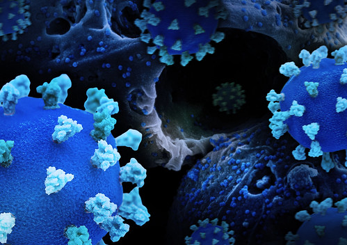
A team of scientists has uncovered a key strategy used by coronaviruses to turn human cells into virus-production factories. Published in a leading scientific journal, the research reveals that during the later stages of infection, viruses like SARS-CoV-2 hijack a specific cellular protein to build large, specialized compartments where new viral particles are assembled.
Previously, it was known that coronaviruses use the cell's internal transport system (the ERGIC and Golgi) for assembly early in infection. However, as infection progresses and these cellular structures break down, it remained unclear where the massive viral assembly required for spreading infection takes place.
This new study provides the answer. The researchers found that several coronaviruses, including SARS-CoV-2 and common cold coronaviruses, trigger the formation of enlarged, vesicle-like structures inside the cell. These compartments are defined by the presence of a host protein called HGS (Hepatocyte growth factor-regulated tyrosine kinase substrate).
Using state-of-the-art imaging techniques—including super-resolution microscopy, electron microscopy, and sophisticated 3D volume imaging (FIB-SEM)—the team directly observed viral structural proteins gathering inside these HGS-positive vesicles. Most importantly, they captured clear images of fully formed and partially assembled coronavirus particles within these compartments.
"We visualized the entire process through cryo-electron tomography," said a senior author of the study. "The viral components are recruited to these HGS-built vesicles, which then serve as the central hub for constructing new virions."
A crucial part of the discovery was demonstrating the importance of HGS. In cells genetically engineered to lack the HGS protein, these specialized virus-assembly vesicles failed to form. While some viruses were still made in other areas, their numbers were drastically reduced, and they were likely defective.
Furthermore, the study revealed that these virus-commandeered "factories" are hybrid organelles, formed by incorporating proteins from the Golgi and the endosomal-lysosomal system. This reorganization creates an optimal environment for the virus to efficiently assemble and then exit the cell, possibly via lysosomal pathways.
The findings build on the team's earlier work identifying HGS as a crucial host factor that physically interacts with the coronavirus Membrane (M) protein. The researchers propose a model where HGS, primarily by binding to the viral M protein, nucleates the formation of the assembly compartment and recruits other viral components to it.
This discovery of HGS's pivotal role opens new avenues for antiviral therapy. Targeting the interaction between HGS and viral proteins could lead to the development of broad-spectrum drugs effective against multiple coronaviruses by disrupting their primary assembly and production site.
This international collaboration was led by teams from the Guangzhou National Laboratory and the Institute of Biophysics, Chinese Academy of Sciences, with key contributions from The First Affiliated Hospital of Guangzhou Medical University. The work utilized advanced core facilities including the Center for Biological Imaging (CBI) in Beijing and the Advanced Bioimaging Core Facility (ABCF) in Guangzhou.
Source: Science China Press